Arteries Diagram / Structure And Function Of Blood Vessels Anatomy And Physiology Ii
Arteries Diagram / Structure And Function Of Blood Vessels Anatomy And Physiology Ii. Chronic lower back pain can have numerous causes, but if you're at risk for clogged arteries, you should consider coronary artery disease a possible cause. Constricted arteries oppose blood flow, and more pressure is required to push blood. The right coronary artery courses in the right atrioventricular groove to. Arteries and arterioles carry oxygenated blood _____ from the heart to the body. Though more often occurring with carotid arteries (the other major ones supplying the brain through the neck), vertebral arteries can be impacted.
ads/bitcoin1.txt
Learn vocabulary, terms, and more with flashcards, games, and other study tools. In fact, about 10 percent of americans have already begun to form blockages in lumbar arteries by the time they're 20. Arteries and arterioles carry oxygenated blood _____ from the heart to the body. The two exceptions are the pulmonary and the umbilical arteries, which carry deoxygenated blood to the organs that oxygenate it (lungs and placenta. Original vintage human anatomy victorian bookplate print 1890s medical diagram veins arteries blood circulatory system of the human body thepapermuseum.
Health Benefits Of Minerals And Vitamins Arteries And Veins Body Diagram Body Anatomy from i.pinimg.com Constricted arteries oppose blood flow, and more pressure is required to push blood. The aorta is the largest artery in the body that exits the left ventricle of the heart. 5 out of 5 stars (293) 293 reviews $ 24.27. Diagram of the human circulatory system (infographic). Arteries and veins are two of the body's main type of blood vessels. The system is responsible for the flow of blood, nutrients. Anatomynote.com found human body artery diagram in detail from plenty of anatomical pictures on the internet. Arteries carry blood away from the heart in two distinct pathways:
The narrowed arteries are at higher risk for complete blockage from a sudden.
ads/bitcoin2.txt
We hope this picture human body artery diagram in detail can help you study and research. The arteries of the head and neck. The arteries of the upper extremity the subclavian artery; For more anatomy content please follow us and visit our website: Circle of willis is indeed a hot neuroanatomy topic! This area is known as the circle of willis. It is a central communication that unites the internal carotid and vertebrobasilar systems. Main branches from the aorta include the brachiocephalic artery, left carotid artery, and the left subclavian artery. The subclavian arteries turn unto the brachial arteries as they pass through the upper arm which feed the radial and ulnar arteries. The cardiovascular system consists of the heart, blood vessels, and the approximately 5 liters of blood that the blood vessels transport. Online quiz to learn arteries of the body; These arteries arise in the neck, and ascend to the cranium. Coronary arteries supply blood to the heart muscle.
Learn the differences between an artery and a vein. These vessels are channels that distribute blood to the body. We think this is the. When the coronary arteries narrow to the point that blood flow to the heart muscle is limited (coronary artery disease), collateral vessels may enlarge and become active. Circle of willis is indeed a hot neuroanatomy topic!
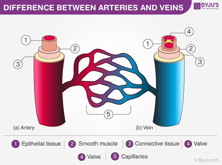
Discover Important Difference Between Arteries And Veins from cdn1.byjus.com Main branches from the aorta include the brachiocephalic artery, left carotid artery, and the left subclavian artery. Classifica'on*of*arteries* • elas'c*arteries* *(conduc'ng*arteries) *aorta,*brachiocephalic,* commoncarod,* subclavian, vertebral,pulmonary,common* iliac. Though more often occurring with carotid arteries (the other major ones supplying the brain through the neck), vertebral arteries can be impacted. We think this is the. Anatomynote.com found blood circulation principal veins and arteries diagram from plenty of anatomical pictures on the internet. Most arteries carry oxygenated blood; These are the only two branches of the ascending aorta. Online quiz to learn arteries of the body;
The anterior tibial artery forms the arcuate artery and its many branches to supply blood to the top of the foot.
ads/bitcoin2.txt
Online quiz to learn arteries of the body; We hope this picture blood circulation principal veins and arteries diagram can help you study and research. Each of these arteries delivers blood to the leg and continues into the foot, with the posterior tibial and fibular arteries forming the plantar arteries and plantar arch that supply blood to the bottom of the foot and toes. An artery (plural arteries) (from greek ἀρτηρία (artēria) 'windpipe, artery') is a blood vessel that takes blood away from the heart to one or more parts of the body (tissues, lungs, brain etc.). Clogged lumbar arteries are far more common than most people realize. For more anatomy content please follow us and visit our website: This allows blood to flow around the blocked artery to another artery nearby or to the same artery past the blockage, protecting the heart tissue from injury. The system is responsible for the flow of blood, nutrients. Most arteries carry oxygenated blood; Diagram of the human circulatory system (infographic). The cardiovascular system consists of the heart, blood vessels, and the approximately 5 liters of blood that the blood vessels transport. The vertebral arteries, and the internal carotid arteries. Within the cranial vault, the terminal branches of these arteries form an anastomotic circle, called the circle of willis.from this circle, branches arise which supply the majority of the.
Coronary arteries supply blood to the heart muscle. Anatomynote.com found human body artery diagram in detail from plenty of anatomical pictures on the internet. The right coronary artery courses in the right atrioventricular groove to. This area is known as the circle of willis. Arteries of the brain and 'circle of willis' diagram.
Coronary Artery Anatomy 3d Anatomy Tutorial Youtube from i.ytimg.com The vertebral arteries, and the internal carotid arteries. The narrowed arteries are at higher risk for complete blockage from a sudden. Blood is pumped from the heart in the arteries. The cardiovascular system consists of the heart, blood vessels, and the approximately 5 liters of blood that the blood vessels transport. Within the cranial vault, the terminal branches of these arteries form an anastomotic circle, called the circle of willis.from this circle, branches arise which supply the majority of the. Anatomynote.com found blood circulation principal veins and arteries diagram from plenty of anatomical pictures on the internet. These arteries arise in the neck, and ascend to the cranium. The typical configuration consists of two coronary arteries, a left coronary artery (lmca) and a right coronary artery (rca), arising from the left posterior and right anterior aortic or coronary sinuses respectively, in the proximal ascending aorta.
Main branches from the aorta include the brachiocephalic artery, left carotid artery, and the left subclavian artery.
ads/bitcoin2.txt
A condition which arises spontaneously or as the result of trauma, where the walls of the artery are split, leading to internal bleeding and disruption of blood flow. This is a list of arteries of the human body. Arteries carry blood away from the heart in two distinct pathways: Circle of willis is indeed a hot neuroanatomy topic! There is a point at which the anterior and posterior arterial circuits of the brain unite or anastomose. We think this is the most useful anatomy picture that you need. Systemic arteries deliver blood to the rest of the body. Over the years, cholesterol plaques can narrow the arteries supplying blood to the heart. Within the cranial vault, the terminal branches of these arteries form an anastomotic circle, called the circle of willis.from this circle, branches arise which supply the majority of the. There are two paired arteries which are responsible for the blood supply to the brain; In fact, about 10 percent of americans have already begun to form blockages in lumbar arteries by the time they're 20. The typical configuration consists of two coronary arteries, a left coronary artery (lmca) and a right coronary artery (rca), arising from the left posterior and right anterior aortic or coronary sinuses respectively, in the proximal ascending aorta. Diagram of the human circulatory system (infographic).
ads/bitcoin3.txt
ads/bitcoin4.txt
ads/bitcoin5.txt
0 Response to "Arteries Diagram / Structure And Function Of Blood Vessels Anatomy And Physiology Ii"


0 Response to "Arteries Diagram / Structure And Function Of Blood Vessels Anatomy And Physiology Ii"
Post a Comment